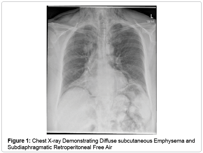
X marfan syndrome (mfs) is a genetic disorder of connective tissue that primarily affects the skeletal, cardiovascular and ocular systems. the development of pneumothorax is common in mfs patients [1], and is often characterized by early onset at a young age and a high frequency of bullae [2-3].
Home Page Journal Of Pediatric Surgery
Best seen with horizontal x-ray beam. upright chest which includes diaphragm, or. upright abdomen which includes diaphragm, or. left lateral decubitus view of the abdomen. patient lies with left side down, right side up. air is seen over the liver. three major findings; free air beneath diaphragm (crescent sign). Barium x-ray 58909; burst, repair of 30403; closure of and repair of musculoaponeurotic layer 45570; closure of, in conjunction with free tissue transfer or breast reconstruction 45569; x-ray, plain 58903; abdominal aortic aneurysm: endovascular repair 33116,33119; abdominal apron: wedge excision 30165,30168,30171.

Johns hopkins medical imaging provides x-ray procedures at convenient locations in green spring station, white marsh, columbia and bethesda. due to interest in the covid-19 vaccines, we are experiencing an extremely high call volume. please. The american heart association explains chest x-rays and answers common questions. a chest x-ray is a picture of the heart, lungs and bones of the chest. a chest x-ray doesn’t show the inside structures of the heart though. retroperitoneal free air x ray a chest x-ray sh. X-rays are a type of radiation called electromagnetic waves. x-ray imaging creates pictures of the inside of your body. x-rays are a type of radiation called electromagnetic waves. x-ray imaging creates pictures of the inside of your body.
Pneumoperitoneum Radiology Reference Article Radiopaedia Org
Detailed information on x-ray, including information on how the procedure is performed due to interest in the covid-19 vaccines, we are experiencing an extremely high call volume. please understand that our phone lines must be clear for urg. Mar 13, 2013 · 1. 0 introduction. x-ray computed tomography (ct) is a well-established tissue imaging technique employed in a variety of research and clinical settings. 1 specifically, ct is a non-invasive clinical diagnostic tool that allows for 3d visual reconstruction and segmentation of tissues of interest. Don't delay your care at mayo clinic featured conditions see our safety precautions in response to covid-19. request an appointment. a chest x-ray helps detect problems with your heart and lungs. the chest x-ray on the left is normal. the i. X-rays use beams of energy that pass through body tissues onto a special film and make a picture. they show pictures of your internal tissues, bones, and organs. bone and metal show up as white on x-rays. x-rays of the belly may be done to.
Xray Computed Tomography Contrast Agents
Mar 24, 2021 · penetrating trauma is an injury caused by a foreign object piercing the skin, which damages the underlying tissues and results in an open wound. the most common causes of such trauma are gunshots a. Aug 30, 2020 · the corticospinal tract controls primary motor activity for the somatic motor system from the neck to the feet. [1] it is the major spinal pathway involved in voluntary movements. the tract begins in the primary motor cortex where the soma of pyramidal neurons are located within cortical layer v. axons for these neurons travel in bundles through the internal capsule, cerebral peduncles, and. Customer service: change of address (except japan): 14700 citicorp drive, bldg. 3, hagerstown, md 21742; phone 800-638-3030; fax 301-223-2400. On conventional radiographs, free air is best demonstrated with the x-ray beam directed parallel to the floor (i. e. a horizontal beam) (see figs. 13-14 and 13-15). small amounts of free air will not be visible on supine radiographs.
What To Expect During A Dental Xray
Mimics of free air: gastric air bubble; a round/oval-shaped air configuration under the left diaphragm. free air is more rind-shaped. on a normal axr, a thick dense (= white) wall can be seen at the cranial side of the gastric air bubble. as opposed to free air, there is a much thinner border between the lungs and the abdomen. More retroperitoneal free air x ray images. In the setting of trauma, if pneumomediastinum is visible on chest x-ray it is termed overt pneumomediastinum whereas if it is only visible on ct then it is termed occult pneumomediastinum 8. pathology etiology. blunt or penetrating chest trauma; secondary to neck, thoracic, or retroperitoneal surgery; esophageal perforation. boerhaave syndrome. Chest x-ray is the first test done to confirm the presence of pleural fluid. the lateral upright chest x-ray should be examined when a pleural effusion is suspected. in an upright x-ray, 75 ml of fluid blunts the posterior costophrenic angle. blunting of the lateral costophrenic angle usually requires about 175 ml but may take as much as 500 ml.
X-ray radiation is used in a variety of places. while it can be helpful, there are also risks. here are five things to know about x-ray radiation. advertisement by: marianne spoon wilhelm roentgen stumbled upon the potential of x-rays while. A chest x-ray looks at the structures and organs in your chest. learn more about how and when chest x-rays are used, as well as risks of the procedure. due to interest in the covid-19 vaccines, we are experiencing an extremely high call vol. As you're sitting in the dentist's chair, you might be told you need a dental x-ray. here's what to expect with this painless procedure and why your dentist may recommend it. Doctors have used x-rays for over a century to see inside the body in order to diagnose a variety of problems, including cancer, fractures, and pneumonia. what can we help you find? enter search terms and tap the search button. both articl.
Notice large amounts of free intraperitoneal air in this abdominal x ray. patient is a 40 year-old female with a ruptured peptic ulcer. www. doctorpaul. org. X current consensus recommendations are to not initiate cervical cancer screening for immunocompetent adolescent females prior retroperitoneal free air x ray to age 21 years. this is in part due to very low rate of 0. 8 per 100,000 new cervical cancer cases diagnosed among women ages 20 to 24 years.
Pneumoperitoneum is the presence of air or gas in the abdominal (peritoneal) cavity. it is usually detected on x-ray, but small amounts of free peritoneal air may be missed and are often retroperitoneal free air x ray detected on computerized tomography (ct). [1] the most common cause of a pneumoperitoneum is a perforation. An erect chest x-ray is probably the most sensitive plain radiograph for the detection of free intraperitoneal gas. if a large volume pneumoperitoneum is present, it may be superimposed over a normally aerated lung with normal lung markings. subdiaphragmatic free gas. leaping dolphin sign. cupola sign (on supine film).
In pneumoretroperitoneum air will collect around the following structures 10: right kidney. referred to as the veiled right kidney sign 7. will not change appearance with patient re-positioning, as opposed to the free air in pneumoperitoneum 11; great vessels. the disappearance of the retroperitoneal inferior vena cava and abdominal aorta 9. Falciform ligament sign (free air). white arrows point to falciform ligament, made visible by a large amount of free air in the peritoneal cavity. the green arrow demonstrate both retroperitoneal free air x ray sides of the wall of the bowel wall (rigler's sign), a sign of free air. the red arrow points to increased lucency over the liver from a large amount of free air. Jun 18, 2021 · abdominal examination was unremarkable except for a painless, soft swelling over the right inguinal region. additionally, his routine blood tests and hydatid titer were within normal limits. chest x-ray (fig. 1) showed absence of a right diaphragmatic shadow, with bowel loops projecting over the lower chest above the level of the liver. a small.
0 Response to "Retroperitoneal Free Air X Ray"
Post a Comment